-
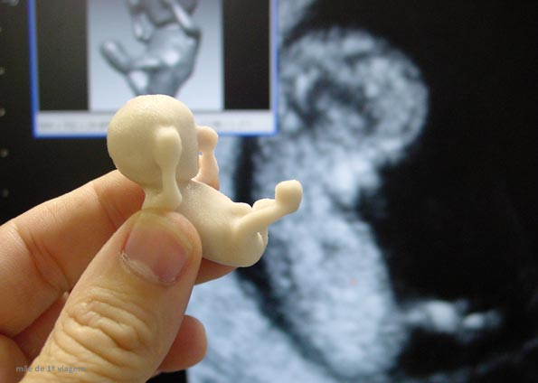
 Beginning of Fetal Development
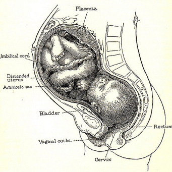
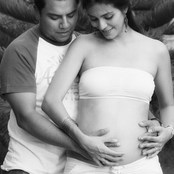
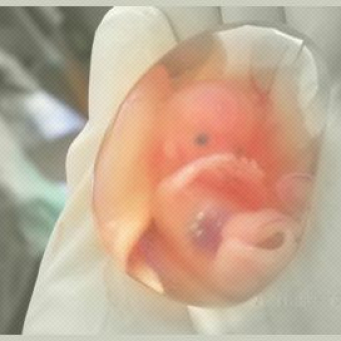
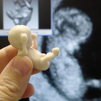
In the third month, the baby develops from an embryo to a fetus as the fetal development stage begins. Although the pregnancy could already be seen through ultrasounds, it is in the coming weeks that the baby starts to acquire the visual features of a human body.
Find the articles in this category that our community has engaged with the most
Your 9th week begins 1-3 weeks... Continue Reading
Your 11th week begins 1-3 weeks... Continue Reading
The long-awaited positive result has finally arrived!... Continue Reading
Your 10th week begins 1-3 weeks... Continue Reading
Your 12th week begins 1-3 weeks... Continue Reading
See the most recent articles published on Famivita
There are several rules that we are used to hearing throughout our lives that do not apply...
Portal hypertension gets its name because it refers to a condition caused by an issue in the...
Conjugal infertility is the term given by the WHO (World Health Organization) for the absence...
The human body is made up of several parts that we often compare to machine gears, which need...
With the advancement of medicine and the ongoing effort to make women’s lives easier and reduce the...
Cardiovascular diseases are at the top of the list of leading causes of death in Brazil, and...
Check out the latest articles published by our medical team in this category