The name can easily be confused with the well-known amniotic fluid, but in reality, it is a condition considered very rare in medicine. Amniotic band syndrome, also known by doctors as amniotic band constriction syndrome, is a little-known occurrence but causes significant concern when diagnosed. That’s why we will explain this condition in more detail, its symptoms, main causes, and the recommended treatments.
What is Amniotic Band Syndrome?
Amniotic band syndrome occurs when pieces and remnants of a tissue similar to the amniotic sac adhere and wrap around the fetus’s limbs during gestational development, forming a band or bandage around the arms, legs, and other parts of the body. As a result, blood cannot circulate properly in these areas of the body, which can lead to malformations and even incomplete development, such as fingers not forming. Depending on where on the body the amniotic band forms, it is even possible for entire limbs not to develop. If it forms in the facial area, it can cause cleft lip or cleft palate. Amniotic band syndrome always occurs in the first trimester of pregnancy, when the fetus is already formed.
Causes of Amniotic Band Syndrome
The causes of amniotic band syndrome are still being studied by medicine, and for this reason, there are only theories that can explain it. The first theory is based on early development in the first trimester, where the baby inside the amniotic sac is in contact with a viscous fluid called the coelom, very similar to egg white. During this period, due to some factors, rupture of this sac may occur, breaking this amniotic membrane (this is not the rupture of the amniotic sac itself, just the membrane), and as the fetus moves, its feet, hands, or any other body part can become “caught in this hole” and get stuck to the body. As gestation progresses and the baby grows, this area that is stuck stops receiving blood properly, resulting in improper development. In some cases, it is possible to observe on an ultrasound some structural parts of the body stuck together, but at this early stage, it is possible that as the fetus moves, it can free itself from what was stuck. The second theory suggests that amniotic band syndrome may be related to bleeding that can occur inside the uterus. If bleeding occurs and the blood clots, it is possible for the baby or a limb to become stuck to this clot, restricting movement and proper development. Another theory is the presence of very strong uterine contractions, which can result in a partial placental abruption. No theories have been found linking amniotic band syndrome to hereditary or genetic factors. However, it has been observed that women who use drugs such as crack, heroin, and cocaine have an increased risk of developing the syndrome.
Is it a Serious Syndrome?
Naturally, receiving such a diagnosis is distressing for parents. But it is important to emphasize that each case must be carefully evaluated to truly understand the severity of the syndrome and which parts were affected or not. The syndrome is quite uncommon, which is why it is considered a rare syndrome, but in general, it does not pose major risks to the baby. The condition can lead to some damage to the baby’s body, such as one less finger or one limb being slightly shorter than the other, but overall, the chances of affecting other body parts more seriously are quite small. As a quick tip for pregnant women: would you like to enhance sexual pleasure during pregnancy? Use water-based lubricating gels that simulate cervical mucus and also balance vaginal pH, reducing the chances of fungus and bacteria responsible for infections proliferating. Famivita has developed the perfect lubricant for women trying to conceive and for pregnant women, FamiGel. You can buy it here in our online store.
How is Amniotic Band Syndrome Diagnosed?
Since it is a gestational event that normally occurs in the first trimester, the diagnosis is made through a morphological ultrasound, and if necessary, a transvaginal ultrasound for further assessment. From the 12th week of pregnancy onwards, it is already possible to detect any occurrences caused by the syndrome and provide more detailed monitoring to anticipate diagnosis. On the doppler ultrasound exam, it is possible to visualize, through a complete body mapping, if there is any compromise in blood flow, even in the smallest parts. 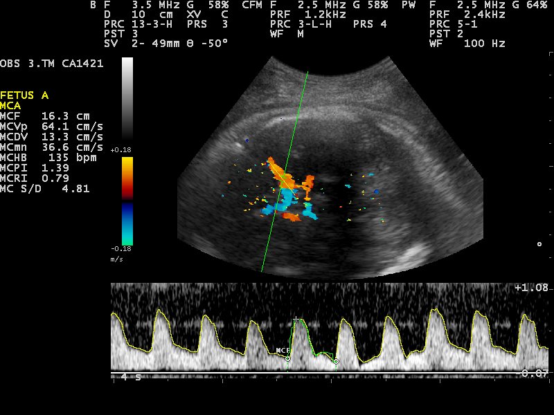
Treatments for Amniotic Band Syndrome
The sooner it is discovered, the lower the risk of complications. This is one of the main reasons why proper prenatal care and especially having a morphological ultrasound in the first trimester is extremely important. Once amniotic band syndrome is diagnosed after the fetus already has a compromised limb, unfortunately, nothing can be done. But when the condition is diagnosed very early in pregnancy, a procedure can be performed to remove the adhesions, allowing the fetus to develop properly. In this type of procedure, the mother needs to receive a spinal anesthesia and be aware that this is a very complex and high-risk procedure for the baby. During the procedure, there are risks of rupturing the amniotic sac, which may lead to pregnancy loss. Due to the high risks, this method is only recommended when it is confirmed that the amniotic band is involving a large part of the fetus’s body, such as an entire arm or a leg. In such cases, the doppler exam is essential to check the blood flow of the involved limb and to study the possibilities and real necessity of the procedure. It is very important to say that not always, when an amniotic band adherence is seen on the fetus’s body, does it mean there will be malformations. Likewise, not every membrane seen during ultrasound means it is amniotic band syndrome. It is very common for specialists to confuse the amniotic band membrane with other residues, for example, remains from another gestational sac if there was a twin pregnancy that did not progress. This is why prenatal care and having all the exams requested by the doctor are essential. In the vast majority of cases, the most recommended treatment is after birth, where malformations are corrected through reconstructive procedures. Only in more severe cases with significant compromise is surgical intervention during pregnancy recommended. Photo: Nevit Dilmen