
The bicornuate uterus, also known as bicornuate uterus, is a congenital malformation of the uterus characterized by a membrane that divides the uterus into two. This membrane can be small or considered complete, totally dividing the uterus into two parts.
This malformation occurs in the uterus during its process of development and organ formation in the fetus, still inside the mother’s womb. A normal uterus is shaped like an upside-down pear and measures around 7.5 cm x 5.0 cm and 2.5 cm thick, but some women have differently shaped uteri. There are many cases where women go their whole lives without ever discovering the fact; in others, they have children without knowing they have an anomaly. Therefore, a bicornuate uterus does not necessarily make pregnancy impossible; it can make it somewhat more difficult, even to carry the baby to 9 months, but there are cases where it is indeed possible to have children—in fact, the great majority of cases.
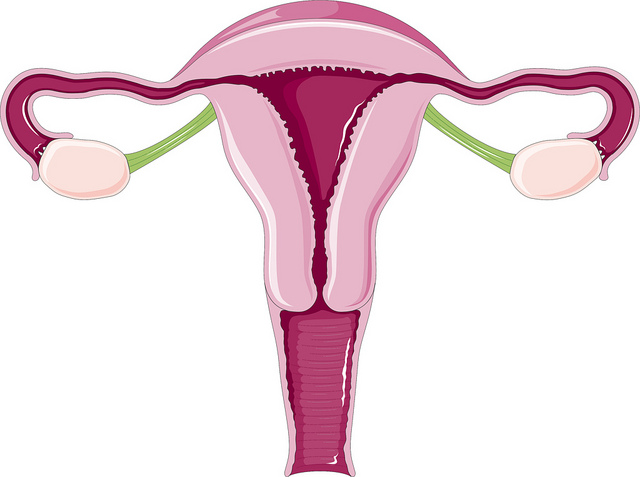
The bicornuate uterus, or in more popular terms, “uterus with two horns” because of its divided appearance, is one of the most common uterine anomalies. It looks like a heart with a slit at the top, which reduces the uterine space and limits room for a developing baby, so the risk of premature birth is high in this scenario. Due to the extra load on the uterus, the cervix can start to open very early, and a cerclage may be needed to hold the baby. Bicornuate uterus can have different degrees. The division of the compartments can be more or less invasive. It is also possible for the divided parts to be of different sizes, meaning they are not always split exactly in half with 100% precision.

Is There Treatment for Bicornuate Uterus?
For women trying to get pregnant who are struggling, a number of tests may be requested by your gynecologist to detect the actual problem and confirm the uterine anomaly, including a simple ultrasound can identify the problem. But specific exams such as hysterosalpingography, an x-ray that uses contrast, can determine the problem more accurately. With it, it is also possible to determine in which part of the uterus it would be most viable for a pregnancy to occur and what the risk is of miscarriage if the embryo settles in the smaller part of the uterus.
Another treatment option is laparoscopy, in which a small cut is made in the abdominal cavity and a device called a laparoscope is inserted, transmitting the image to a monitor. During the procedure, the doctor may be able to remove a septum that is blocking the passage. Removing the septum will give the uterus its usual appearance and then, the chances of miscarriage become much lower.
Surgical treatment for bicornuate uterus is only indicated when it prevents or seriously hinders pregnancy. However, since these are isolated cases, the doctor will conduct a thorough analysis to assess which cases require greater attention, and which may not require surgery at all. I have seen many women with bicornuate uterus become mothers without any problem; it just takes a bit of luck and a lot of faith.


