At the beginning of pregnancy, some structures are created. They are not part of the embryo’s body, but are there to assist in its vital functions. This is the case with the yolk sac, which, along with other structures, creates the conditions for the baby’s development. 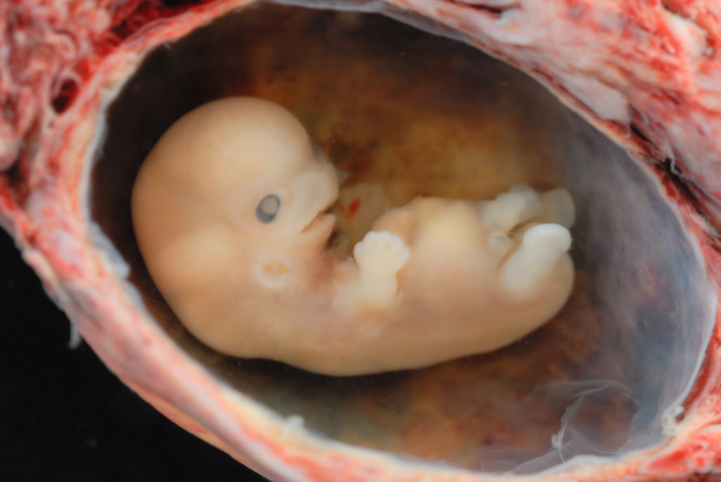
What Is the Yolk Sac?
The yolk sac (or vitelline sac) is the first element observed in the gestational sac during pregnancy, usually from the 3rd day of gestation. It does not function for long, about three months, but it is very important for fetal development1. Much of it will be incorporated into the primordial intestine from the 4th week of gestation. This is why the size of the yolk sac during the first six weeks after the egg is fertilized is much larger than the size of the amniotic cavity, along with the emerging embryo.
Why Is a Yolk Sac Needed?
Each stage of pregnancy has characteristics that are quite reliable indicators that the pregnancy is healthy and developing as it should. In the early stages of development, when the embryo is forming, only the yolk sac plays a variety of roles, without which the baby’s normal development would be impossible.
Functions of the Yolk Sac
- The yolk sac acts to provide nourishment to the embryo during the initial stages until the placenta takes over, which happens around the 8th week of pregnancy. It also acts as a system that allows blood and nutrients to circulate throughout the embryo, called the circulatory system.
- From the 18th day after the start of a new life in the yolk sac, the first embryonic red blood cells (erythroblasts) are formed and capillaries begin to grow, after which the entire fetal circulatory system is formed2.
- From the 28th day, the walls of the yolk sac produce the first germ cells, which will later migrate to the embryonic gonads. It is important to note that, at this stage, the eggs of a future girl are formed. If, at this moment, the mother has suffered or is suffering severe stress – egg formation may occur incorrectly and, in the future, an adult woman may suffer from infertility.
- Up to the 6th week, the yolk sac acts as a “primary liver” and produces proteins that are very important for embryonic development, such as alpha-fetoprotein.
The yolk sac is also actively involved in metabolic processes, the formation of immunity, and helps replenish fetal excretions.
Can You See the Yolk Sac on Ultrasound?
On ultrasound, the yolk sac is visible from the 5th to the 12th week of pregnancy. If the doctor does not see the yolk sac, this condition is considered unfavorable as it may be a sign that the pregnancy could stop developing at any time. Premature disappearance of the yolk sac is also an unfavorable sign. Normally, the yolk sac should have the following dimensions:
- Less than 5.5 mm at 5–10 weeks;
- More than 2 mm at 8–12 weeks.
This happens because the diameter of the yolk sac increases exponentially between the 5th and 11th weeks of pregnancy, decreasing soon after, in accordance with fetal growth. At the end of the first trimester, the fetus is fully formed and moves on to the placental circulatory type. The yolk sac becomes redundant and is reduced so that it can no longer be seen on ultrasound. Until the end of pregnancy, it remains as a small, somewhat oval body, with a diameter varying from 1 mm to 5 mm at the base of the umbilical cord. If the yolk sac is reduced before the right time, that is, when all organ systems in the fetus are not yet formed, the pregnancy will not develop properly. The embryo will not be able to progress to a new stage of development and will not become a fetus. This often leads to a non-viable pregnancy and miscarriage.
What Happens If the Yolk Sac Is Not Visible?
If the yolk sac is not visible between the 5th and 11th weeks of gestation, there are several possibilities to consider:
- Calculation error of conception – The pregnancy may not have been properly identified and is less than 6 weeks old (up to 6 weeks, the yolk sac is usually not visible).
- The ultrasound may detect the embryo’s heartbeat (7 weeks) and the embryo is visible. But if no yolk sac is seen after 7 weeks, it may result in interruption of the pregnancy.
- Unfavorable prognosis – The woman should have a new ultrasound, preferably a high-resolution transvaginal one, to check for the presence or absence of the yolk sac.
- After the 12th week, if the yolk sac is not identified, it usually means the fetus has started receiving nutrients through the placenta.
What Are Embryonic Annexes?
Just like the yolk sac, embryonic annexes are temporary structures that are created to provide the baby with the right conditions for development during its time in the womb. They are:
- Yolk sac;
- Amniotic sac or amnion;
- Allantois;
- Chorion; and
- Placenta.
What Does Irregular Gestational Sac Mean?
During the first trimester, one of the items checked about the gestational sac is its contour, since if the shape is irregular, the risk of sac detachment and miscarriage is greater3. In these cases, the pregnant woman should remain at rest and follow the medications prescribed by the doctor so the gestational sac returns to normal and the pregnancy develops properly. See also: Gestational Sac Detachment – How to Detect and Treat Photos: lunar caustic